

Five talks in a Sunday morning session showed how thinking about diabetic eye disease is shifting, from protecting the whole retinal neurovascular unit to using AI for screening, and from a single view of macular oedema to four distinct phenotypes.
Diabetic retinopathy has long been seen as a microvascular complication. A session on the morning of Sunday 4 October, chaired by Edoardo Midena and Elisabetta Pilotto (both Padova, Italy), made the case that this view is no longer enough, and showed what comes after it.
From retinopathy to retinal disease
Professor Tim Curtis (Queen’s University Belfast) opened by describing the retinal neurovascular unit, the interdependent group of neurons, glial cells, pericytes and endothelial cells that keeps the retina in balance. In diabetes, he explained, every cell type is affected. Neuronal dysfunction can come before vascular dysfunction, and because the cells are so interconnected, protecting the neurons and glia protects the vessels, and the other way round.
During discussion, a delegate noted that major recent reviews now use “diabetic retinal disease” (DRD) in place of “diabetic retinopathy”, because the older term keeps the focus on blood vessels alone. Curtis said his group has been doing the same.
A new target: acrolein
His group has focused on acrolein, a highly reactive aldehyde that builds up in the diabetic retina and forms protein adducts, concentrated in Müller glia and neuronal layers, in both rat and human tissue. They identified a potent acrolein scavenger, 2-HDP, and tested it in diabetic rats at 100 mg/L in drinking water for up to six months, in work funded by Diabetes UK. It had no effect on blood glucose, weight or blood pressure, yet it reduced the adducts in Müller glia, preserved the electroretinogram, prevented loss of synaptic proteins and retinal thinning, reduced vascular leakage, prevented pericyte dropout and the formation of acellular capillaries, and dampened inflammatory signalling, including cytokines such as ICAM-1 and MCP-1. Curtis suggested acrolein scavenging could be a translational strategy for early diabetic retinal disease.
Retinal tests for cognitive impairment
Professor Noemi Lois (Queen’s University Belfast) presented results from the RECOGNISED consortium, funded under the EU’s Horizon 2020 programme and co-led with Rafael Simó. People with type 2 diabetes face a higher risk of mild cognitive impairment, which affects self-management and raises the risk of glucose extremes and hospital admission, yet there are few tools to detect it in clinic.
Among 313 participants, 128 had normal cognition and 185 had mild cognitive impairment. Those with impairment had lower retinal sensitivity, less stable fixation, a higher pupillary area ratio and a lower photopic B-wave amplitude. OCT measures did not separate the groups. The best model, combining microperimetry, a handheld RETeval electrophysiology test, years of education, a diabetes-specific dementia risk score and the MoCA, reached an area under the curve of 0.84, with sensitivity of 79.9% and specificity of 79.0%. A simpler version without microperimetry still performed well. HbA1c did not distinguish the groups. The aim is to add such tests to diabetes screening so patients who need cognitive support can be found earlier.
AI for screening, prediction and beyond
Professor Tien Yin Wong (Tsinghua Medicine, Beijing, and Singapore National Eye Centre) reviewed how far AI has come for diabetic retinopathy screening. His message on detection: you can’t simply buy a model off the shelf. Performance depends on the local population, which is why Singapore developed SELENA+ for its Chinese, Indian, Malay and Caucasian groups. Economic modelling also showed that AI does not produce large savings, because the infrastructure is expensive. Foundation models, trained on 1.6 million fundus and OCT images and then fine-tuned for individual tasks, can detect many conditions, but he cautioned that a broad model may be less specific than one built for local needs.
On prediction, models that estimate future risk could stretch the average screening interval from 12 months to about three years. In the study he described, around 30% of patients would still need yearly screening, while about 17% could safely go to five years. He also described “oculomics”, using a fundus photograph as a window on the rest of the body, with estimates of blood pressure, HbA1c, kidney function and coronary calcium, and opportunistic screening for cognitive impairment. His caution was that external validation is increasingly difficult, so models now have to be validated locally.
Hypertension and retinal ischaemic lesions
Amani Fawzi (Cole Eye Institute, Cleveland Clinic) turned to the patient in front of the clinician. Hypertension coexists with diabetes in about 80% of type 2 and 30% of type 1 patients, and it triples or more the risk of progression to proliferative disease. Her group studied retinal ischaemic perivascular lesions, known as RIPLs or “ripples”: atrophic scars left on OCT by old ischaemic infarcts.
In 129 eyes, the mean RIPL count rose from 1.2 in diabetes without retinopathy to 3.7 in non-proliferative and 10.1 in proliferative disease, and the share of patients with hypertension rose from 41% to 76%. More than 2.5 RIPLs flagged a high risk of vision-threatening retinopathy, with an area under the curve of about 0.8, validated on a second device and population. The RIPL burden may explain about 30% of hypertension’s effect on retinopathy. Eyes with referable disease and hypertension also had more ischaemia in the deep capillary plexus, and that ischaemia correlated with vision only in the hypertensive eyes.
Fawzi now counts RIPLs before she sees the patient. Three or more would prompt her to order fluorescein angiography. The work excluded eyes with macular oedema, where cystoid change would probably hide the lesions.
One size does not fit all in DME
The session closed with Edoardo Midena (University of Padova), speaking on behalf of the AI in DME study group of the Italian Retina Society. His argument was that diabetic macular oedema is not one disease. Around 40% of patients treated with anti-VEGF do not improve, as shown in the 2016 analysis of Protocol T and in a recent Swiss real-world study. Choosing a treatment, he said, is sometimes “more or less flipping the coin”. Central retinal thickness correlates poorly with vision, but OCT contains far more information: inflammatory hyperreflective foci, disruption of the external limiting membrane and ellipsoid zone, and the distribution of fluid.
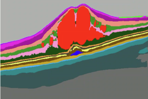
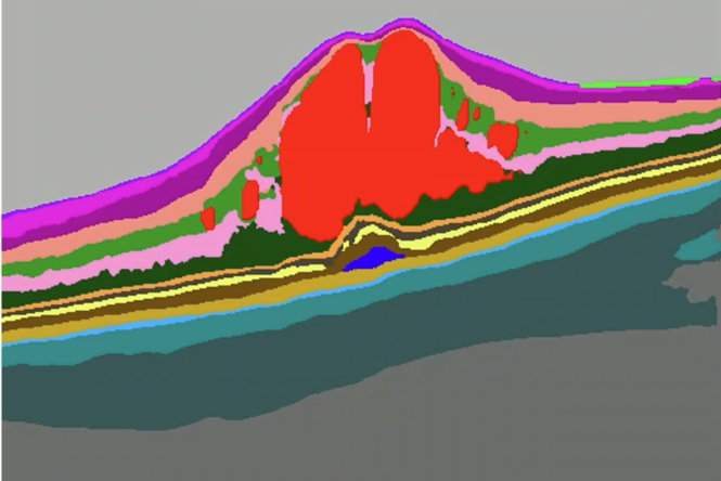
He applied a CE-marked AI algorithm to 2,355 eyes of 1,688 patients. Unsupervised clustering found four phenotypes, differing in intraretinal fluid volume, subretinal fluid and outer-retinal disruption. Phenotypes 3 and 4, with subretinal fluid, outer-retinal changes and more inflammatory foci, made up about 40% of treated eyes. In 404 untreated eyes the same four phenotypes appeared, with phenotypes 3 and 4 accounting for about 60%. Ellipsoid zone disruption was more common than external limiting membrane disruption (in 38% of eyes against 22%). The external limiting membrane, a marker of Müller cell activity, emerged as a key driver, linked to subretinal fluid and to visual function.
His conclusion slide was blunt: DME needs personalised treatment based on “precision medicine”, not a one-size-fits-all approach.