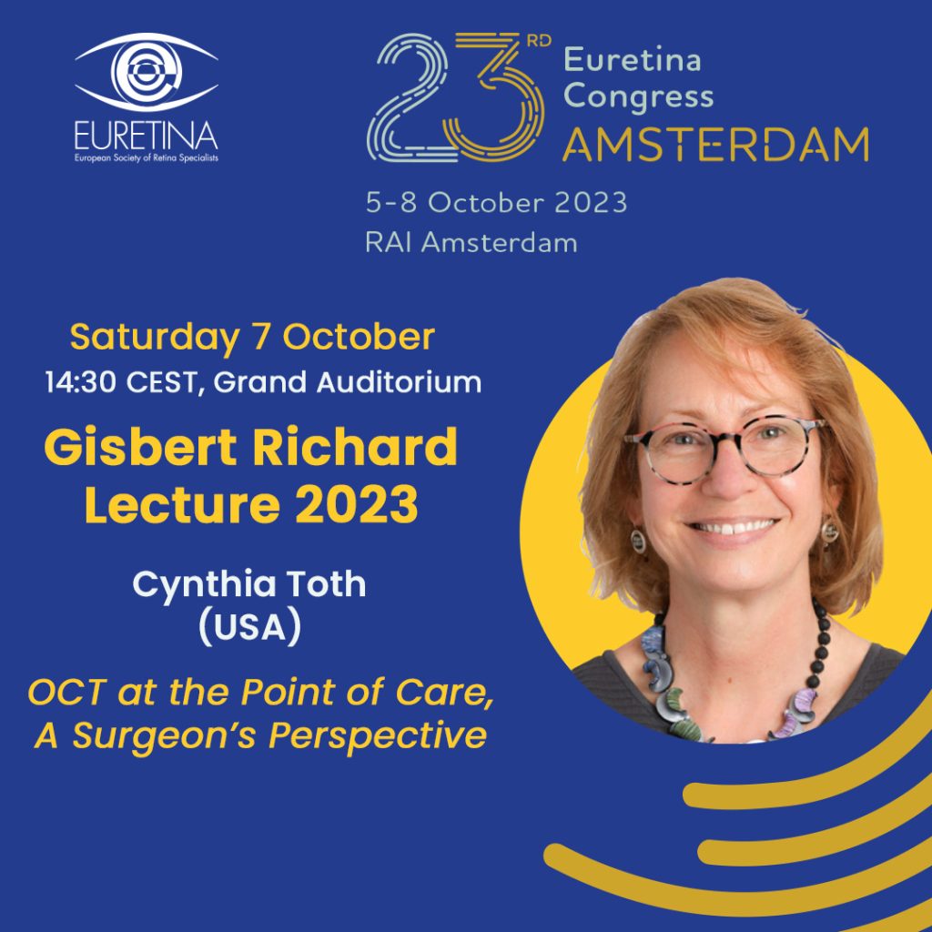
Gisbert Richard lecturer describes her 20+ year collaboration with engineering, computer science, and clinical colleagues and their ongoing journey to improve eye care through innovations in handheld and intraoperative OCT
Without exclusion, retina specialists recognise the value brought by optical coherence tomography (OCT) to the care of patients with retinal disease. However, having access to the technology only by sending patients to the photography area of the clinic was frustrating for physicians and surgeons who could benefit from having clinically relevant OCT data at the point-of-care, said Dr Cynthia A Toth, Professor of Ophthalmology and Biomedical Engineering, Duke University School of Medicine, USA. In her Gisbert Richard Lecture, Dr Toth will share the pioneering work done with valued collaborators to address that need by creating OCT systems that serve as practical tools for paediatric and surgical applications.
She will present a timeline documenting the evolution of point-of-care OCT for retina specialists that starts approximately 30 years ago when Dr Toth crossed paths with Joe Izatt, PhD, a biomedical engineer working on developing OCT for ophthalmology.
“Once Dr Izatt joined the faculty at Duke, the development of point-of-care OCT exploded. The ability to partner with an engineer able to build the hardware for systems that could meet our clinical needs was invaluable. At the same time, input from clinician colleagues and the expertise of computer science engineer Sina Farsiu, PhD, who came up with solutions for generating relevant clinical information through image analysis has been critical to our success creating practical point-of-care OCT technology.”
Dr Toth will share the work done to develop handheld OCT for examining paediatric patients at the bedside and the advances that have occurred with intraoperative OCT since it was introduced in 2010.
“The first challenge for bringing OCT into the OR was to develop a compact unit that could attach to the surgical microscope. The second challenge was to address the need for faster imaging speed,” she said.
Dr Toth will talk about available commercial systems for intraoperative OCT and present cases showcasing some applications. She will also describe the latest generation of intraoperative OCT – real-time volumetric (“4D”) swept-source OCT – that is being used at Duke to image patients through an institutional review board approved protocol.
Dr Toth will conclude with the exciting research underway using image fusion to merge the microscope and OCT views into a single image and discuss its implications for improving surgery and patient outcomes through guidance during complex surgeries and integration with robotic systems for increasing surgical precision.
Dr Toth will close with an important message for her colleagues. “So that these new systems will help us improve our surgery and its outcomes, we as clinicians need to communicate with the engineers and companies developing the technology and be specific about what is relevant for us in real time. It is not enough to just say, ‘we want something better’,” she said.
Dr Toth will deliver the Gisbert Richard Lecture at 14:30 on Saturday 7 October 2023 in the Grand Auditorium.


