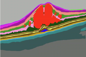


The Euretina Session track at the 2026 Congress began on Thursday morning with a programme titled “Eye Openers”. In this video-based interactive session, six experienced retina specialists presented lessons learned from challenging vitreoretinal surgery cases. At junctures when complications occurred throughout the cases, input on next steps was sought from a faculty panel and audience members equipped with electronic response keypads.
Leading off the session, José García-Arumí (Spain) featured a case involving a 77-year-old man with a history of four unsuccessful surgeries for retinal detachment now presenting with extensive subretinal proliferation involving the macula, hand motion visual acuity, and inferior retina contraction related to hypotony (IOP 4 mmHg). Further complicating the management decision was the fact that the gentleman had a back injury causing difficulty maintaining a face-down position.
Dr. García-Arumí described his approach for removing the proliferative tissue while minimising photoreceptor damage. More complications were encountered after subretinal traction but were managed successfully. Providing key messages, Dr Arumi said that in extensive subretinal PVR with inferior retina contraction and macular involvement, a larger retinotomy is better than surgery using small incisions. He also noted that silicone oil is a good tamponade option in patients who have difficulty maintaining a prone position.
In a case subtitled “How to convert a very easy case to a very tough one”, Stratos Gotzaridis, MD (Greece) showed 1) why a plan to take shortcuts may not be a good idea, and 2) persistence pays off, even when confronted with a series of complications. “Never give up. Remember the sun always comes out after the storm,” Dr Gotzardis said. The case involved a 78-year-old gentleman with a dislocated SUNSET IOL and a history of vitrectomy for retina detachment. Extenuating circumstances prevented Dr. Gotzaridis from proceeding with his preferred approach to implant a Carlevale IOL. So, he aimed instead to follow a minimal surgical plan in which he would scleral fixate the existing IOL rather than exchange it. Several complications later, the IOL was exchanged for a three-piece MA60. At the 1-month follow-up, the patient had a good refractive and visual outcome with no evidence of cystoid macular oedema.
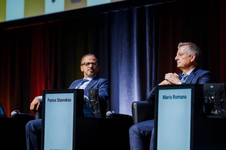
 In the next presentation, Mario Romano, MD (Italy) shared a case involving repair of a giant retinal tear with management of retinal slippage that occurred during BSS-air exchange. His approach for optimising PFCL-air exchange and thus retinal flattening was a key point in his presentation. He emphasised the importance of recognising the change of interfacial tension at the PFCL bubble-air interface and its effect on the shape of the PFCL bubble once no BSS remains. “A well-performed PFCL-air exchange can allow gas tamponade even in giant retinal tears,” Dr. Romano said.
 Panos Stavrakas, MD (Greece) presented a patient with a rhegmatogenous retinal detachment, extensive preretinal and subretinal proliferative vitreoretinopathy (PVR), and inferior retinal thinning. The subretinal PVR presented as both a wide layer underneath the retina and with the typical subretinal bands. Additionally, a wide sheath of subretinal PVR had formed underneath the macula causing a fixed fold. Dr. Stavrakas said that retinectomy with a bimanual technique was the only way to remove the PVR since an external approach would probably not have been successful. He pointed out the need for repeated PVR peeling even if there is no staining and for recognising indirect signs for presence of PVR.“It is important to try to guess at the beginning of the case what you might encounter. Sometimes the decisions you make at the start can make a lot of difference in the end for our patient’s vision,” he said.
Lejla Vajzovic, MD (United States) shared a rare scenario encountered during primary phakic retinal detachment repair. The patient was a 62-year-old male presenting with a three-day history of blurry vision in his right eye who was found to have a macula-off retinal detachment. The repair was undertaken under peribulbar anaesthesia. During PFCL injection it was noticed that the bullous area of subretinal fluid suddenly disappeared without being aspirated. Scleral infolding was noted as well. The decision was made to stop and check the infusion. External exploration performed after confirming the infusion cannula had not been dislodged revealed an area of staphyloma in the superotemporal quadrant and a rupture at that site that was managed with a primary closure. The case proceeded with laser exchange of the PFCL and tamponade with long-acting gas. The patient did well. At 1 year postop the retina was attached, IOP was 13 mmHg, and VA cc was 20/80.
In the last presentation of the session involving a case of retinal detachment repair, Ehab El Rayes, MD, PhD (Egypt) highlighted the challenge posed by the presence of subretinal strands. He noted that although not all subretinal strands need to be removed, when indicated, removal should be done safely to avoid complications. His video demonstrated easy ways for removing the strands as well as mistakes and how to avoid complications.
All sessions are available to view on demand for registered attendees.