

Euretina lecturer discusses pathologic myopia with a focus on its vision-threatening complications and the insights gained from their characterization through the application of advanced imaging technologies
Pathologic myopia is a major cause of blindness, although many patients with pathologic myopia maintain relatively good corrected vision. In her Euretina Lecture, Dr Kyoko Ohno-Matsui, MD, PhD will discuss the complications of pathologic myopia that lead to significant vision loss and the importance of findings from the use of three-dimensional magnetic resonance imaging (3D MRI), ultra-widefield optical coherence tomography (UWF-OCT), and polarization-sensitive OCT (PS-OCT) to study eyes with pathologic myopia.
The complications of pathologic myopia that lead to significant vision loss include myopic macular neovascularization (myopic MNV) and MNV-related macular atrophy, myopic traction maculopathy, and optic nerve damage. Speaking about these complications, Dr Ohno-Matsui will discuss a study conducted using machine learning models that identified prior myopic MNV and category 4 macular atrophy (mostly after MNV) as two of the four most important variables for predicting long-term visual acuity in highly myopic eyes. In addition, she will present findings from imaging studies and their implications.
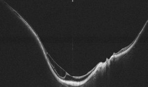
“Using UWF-OCT we identified intense vitreous traction on the retinal vessels in eyes with myopic traction maculopathy, suggesting that retinal vessel traction may be more significant than foveal traction,” Dr Ohno-Matsui said.
In the remainder of her lecture, Dr Ohno-Matsui will focus on posterior staphyloma, which is considered the hallmark lesion of pathologic myopia and the underlying cause for its vision-threatening complications. She will discuss the importance of using 3D MRI and UWF-OCT for visualizing the full extent of staphylomas and how the findings from research using those technologies have resulted in rethinking of concepts about staphyloma formation and is facilitating understanding of the mechanisms for damage to visually important tissues in eyes with pathologic myopia.

Acknowledging the role of 3D MRI as a powerful tool for research studies relating to pathologic myopia, Dr Ohno-Matsui will note the value that UWF-OCT offers for screening purposes and how such use may provide understanding of when, where, and how staphylomas develop.
“Identifying the timing, location, and specific tissues where staphyloma originates can lead to treatments aimed at preventing or controlling its progression before sight-threatening complications occur,” she said.
In the last portion of her lecture, Dr Ohno-Matsui will discuss how PS-OCT is also being used to gain an understanding of staphyloma pathogenesis.
“When we think about staphyloma, the sclera is obviously the most important target tissue, and Dr Nevile McBrien has suggested that the orientation of scleral collagen fibers has a role in staphyloma pathogenesis. Imaging with PS-OCT allows us to visualize the density and orientation of the collagen fibers,” she said.
Professor Ohno-Matsui will deliver the Euretina Lecture in the Grand Auditorium during the Opening Ceremony at 16:35 CEST on Thursday 19 September.


")
")
")