

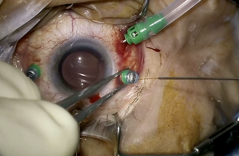
Four teams showed where robots stand in vitreoretinal surgery today, from first-in-human trials to a subretinal injection performed across 4,200 kilometres. The shift under way is from single devices to AI-assisted surgical ecosystems.
Robots in retinal surgery are no longer a concept. In a session chaired by Nicole Eter and Peter Stalmans, speakers presented clinical results from several platforms and looked ahead to what comes next.
Why robots, and what they do
Introducing the session, Peter Stalmans set out the problem. Surgeons are limited by tremor, fatigue and a lack of force feedback, and those limits matter more as treatments move into very small spaces. A robot offers three core capabilities. Tremor filtering removes high-frequency hand movement. Motion scaling translates a large movement into a small one, for example a 10:1 reduction. Position locking holds an instrument still at the chosen spot. A remote centre of motion makes the instrument pivot through the sclerotomy, which stabilises the eye. The two main applications he highlighted were retinal vein cannulation with thrombolysis and subretinal delivery of drugs and gene therapy.
Subretinal injection with the Mynutia system
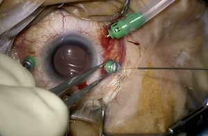
Ivo De Clerck (UZ Leuven) presented the Lotus trial of the Mynutia system, a co-manipulation robot in which the surgeon holds the handle directly while the system cuts tremor tenfold and can freeze the instrument. Because gene therapy patients are scarce, the proof-of-concept study used submacular haemorrhage, injecting through the retina next to the blood.
The primary endpoints were met in full. In all 20 eyes the cannula entered the subretinal space and stayed there for at least three minutes, cannula removal was safe in every case, and there were no inadvertent retinal touches and no device deficiencies or robot-related adverse events. The retinotomy closed spontaneously after the needle was removed, even after three to five minutes. Blood displacement was complete in 95% of eyes, the lowest pressures were 0.3 bar to initiate and 0.1 bar to propagate, and vision improved by more than three lines in two-thirds of patients. In one case the surgeon had to go directly through the haemorrhage because the system’s range was too limited.
De Clerck was candid about the limits. With tremor removed, human depth perception is now the limiting factor, blood is hard to see in real time at such low pressures, the current version only approaches from the temporal side, and a haemorrhage model does not perfectly stand in for gene therapy. His answer is intraoperative OCT with AI-driven real-time guidance.
The Preceyes system: from the macula to retinal veins
Jasmina Cehajic-Kapetanovic (University of Oxford) described the Preceyes system, a teleoperated robot with a joystick, motion scaling, virtual boundaries and a clutch that freezes the position. In the first-in-human R2D2 trial, it was used for macular hole surgery and for subretinal TPA in wet AMD, under local anaesthesia, with no adverse events in either arm. Haemorrhage resolved at one month, as in the control arm. A systematic review has since found 12 clinical studies, three of them randomised trials, with no concerns about ocular harm.
Her team is now focusing on retinal vein occlusion, the second most common cause of permanent sight loss in the UK. A third of cases lose vision despite optimal long-term treatment, and a third of ischaemic cases go on to neovascular glaucoma and painful blind eyes. Injections treat the complications rather than the clot, and the veins are too small for manual cannulation. In silicone-model simulations, 117 of 122 attempts succeeded (96%). Automated needle tracking showed significantly less tremor with the robot, with a tremor radius of around 90 microns, against vessels of 150 to 200 microns. Stainless-steel microneedles penetrated better than the Teflon needles used in subretinal surgery. After successful work in a primate model, an ongoing randomised trial has begun, with first human surgeries performed. At one week the first patient showed reduced macular oedema.
A map of the field, and telesurgery
Koorosh Faridpooya (The Rotterdam Eye Hospital) argued that “robotic device” is the wrong term now: the field is moving towards a robotic ecosystem that links preoperative data, the robot and postoperative analysis. He surveyed the active platforms. They include the Mynutia, the bimanual LUCA, two AI-assisted cataract robots (Foresight Robotics and the Horizon system from UC) with no retina use yet, and a Norwegian bimanual system that has demonstrated telesurgery but not yet entered human trials.
He then described the Chinese Oculotronics platform, founded in Guangzhou in 2017, with 10-micron precision, AI guidance and a three-dimensional force sensor. It has been used in clinical studies at more than 11 centres, and a regulatory submission is under way. In June 2023 the company carried out the first remote micron-level animal eye surgery across the sea between Hainan and Guangzhou. In November 2025 it completed the first human remote robotic subretinal injection, across 4,200 km between Guangzhou and Urumqi, for a 42-year-old man with submacular haemorrhage. The haemorrhage had resolved by day 23 with no adverse events. Latency was the central challenge. The team used public and private 5G networks with its own communication layer, and trains surgeons in a digital-twin simulator.
Bimanual surgery with LUCA
Fanny Nerinckx (Ghent University Hospital and Chirec Delta Hospital Brussels) closed the talks with the LUCA system from Acusurgical. Its two independent arms are driven from a piloting station, and it fits into an existing operating room using standard disposable instruments. In the first-in-human trial of seven patients (six epiretinal membranes and one macular hole), there were no device-related adverse events and no iatrogenic retinal tears. Surgery was completed in every case, and gesture fidelity was rated “very satisfactory” in 100% of them. She also noted that the surgeon sits more comfortably than at a conventional microscope.
A second version is planned with patient-movement detection, virtual boundaries, automation of some steps and surgical analytics, and a registration study is designed as a randomised two-arm trial. The aim, she said, is “a full intelligent AI-assisted surgical suite”.
What comes next
Across the talks the direction was the same: more precision, then more intelligence. Speakers pointed to broader trials in vein and arterial occlusions, earlier treatment windows, better visualisation and workflow integration. Cost-effectiveness is still to be shown. A panel discussion closed the session.