

Speakers in the Saturday morning Euretina Session on myopia covered topics relating to myopic traction maculopathy (MTM), myopia control in children, and dome-shaped macula.
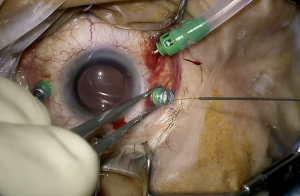
In the first talk on MTM, Tomonari Takahashi, MD (Japan) reviewed advanced imaging techniques and presented data from a study characterising long-term outcomes. Noting that use of appropriate imaging modalities is essential for evaluating retinal changes in eyes with MTM to determine indications for surgery and for postoperative follow-up, he discussed the utilities of ultra-widefield optical coherence tomography (OCT), retromode imaging, and DREAM OCT, a new system from Intalight.
The long-term follow-up study showed that fovea-sparing internal limiting membrane peeling may have a protective effect against the postoperative development of macular holes. In addition, despite certain considerations, early intervention was associated with favorable surgical outcomes. Dr Takahashi emphasised the importance of carefully evaluating changes in the peripheral retina as well as in the posterior pole postoperatively and the need for extended monitoring over an extended period because of the potential for adverse events developing during long-term follow-up.
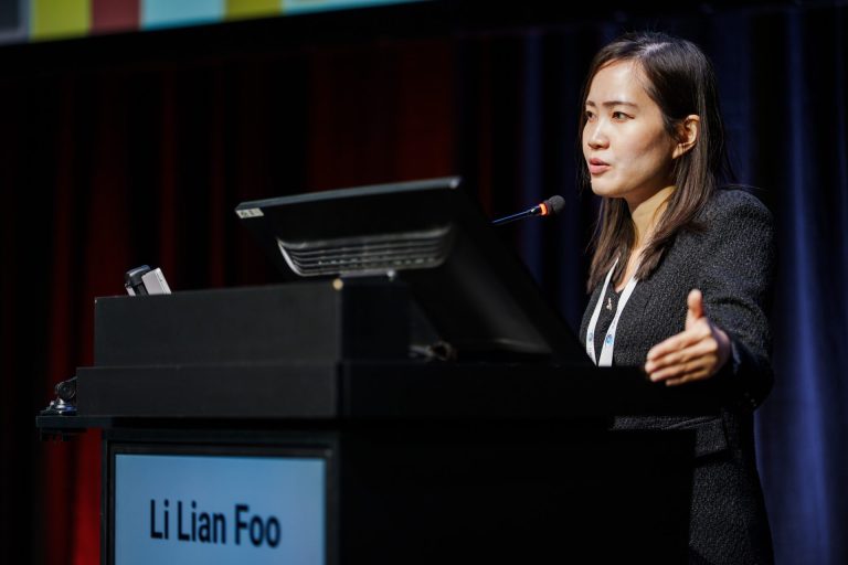
Li Lian Foo, MD, PhD (Singapore) covered the use of atropine for preventing high myopia. Her presentation described the early clinical trial experience in Asia with low-dose atropine to the new era of regulated pharmacological myopia control in Europe with the approval of atropine sulfate 0.1 mg/mL (Ryjunea) to slow the progression of myopia in children and adolescents. She reviewed the STAR trial that provided evidence about the efficacy of atropine in a predominantly Caucasian population and discussed Consensus Guidelines on Management of Childhood Myopia in Singapore, highlighting information about the use of atropine, including when and how it should be started and when and how to stop it.
Matteo Mario Carlà, MD (France) presented findings about the longitudinal progression of myopic maculopathy from a retrospective single-centre study of a large European cohort followed at the Instituto de Microcirugía Ocular in Barcelona for a mean of more than 11 years. He reported that 57% of eyes progressed during follow-up, patchy atrophy was the main gateway to macular atrophy, and compound (type IX) staphyloma carried the highest risk.
Dr. Carlà also presented findings from new analyses of the cohort showing that subfoveal choroidal thickness declined fastest in eyes with diffuse atrophy and that a choroid thinner than 40 µm predicted macular atrophy and visual loss. Additionally, the new analyses showed that perivascular patchy atrophy affected more than 50% of eyes, progressed in 90% and coalesced into linear atrophy, and presence of three or more arcades was associated with high risk of foveal atrophy. Summing up the clinical implications of the findings, Dr. Carlà said that atrophic stage, choroidal thickness and the extent of perivascular atrophy together provide a practical risk profile that can help clinicians identify the highly myopic patients who need the closest follow-up.
Elise Philippakis, MD (France) presented findings about the prevalence, morphology and outcomes of dome-shaped macula (DSM) in eyes with high myopia in the first large European cohort. The research was performed using data collected at the French Myopia Institute. She reported that the study confirms the prevalence of DSM in highly myopic adults and children and showed a horizontal axis predominance. Comparing based on analyses of data from the first large European cohort French Myopia Institute cohort. She noted that the findings in this large European cohort confirm the prevalence of dome shape macula in highly myopic eyes but also in paediatric patients with predominance of the horizontal axis configuration. Risk factors for macular curvature changes were axial length in both children and adults and possibly greater astigmatism. Discussing findings on complications, Dr Philipakis said that the rate of serous retinal detachment was relatively low (6%), possibly because the French Myopia Institute is not a tertiary center, and dome height > 300 µm was strongly associated with its prevalence. Choroidal neovascularisation was seen in one-third of cases and was strongly associated with macular atrophy. As her take-home messages, Dr. Philipakis advised screening early with routine SD-OCT for all paediatric eyes with axial length >25.0 mm to detect early scleral curvature changes, treating myopia aggressively in children to control axial elongation, and to continue longitudinal OCT surveillance during transition to adulthood and inform patients regarding complications of DSM.
The session concluded with co-moderators José M Ruiz-Moreno, MD, PhD (Spain) and Kyoko Ohno-Matsui, MD, PhD (Japan) covering surgical controversies in myopic traction maculopathy. In this interactive format integrated audience participation with response keypads, the two speakers presented a series of cases with questions interspersed asking attendees whether or not they would operate and to indicate their preferred surgical technique.